Digital Radiography
At Complete Dental Care of Avon we keep pace with modern dental practices, and we have invested in a state-of-the art, low radiation digital x-ray system. This advanced technology offers increased safety by reducing our patients exposure to radiation while allowing us to increase our diagnostic capabilities immensely.
Digital radiography utilizes computerized equipment and software to capture x-ray images of your mouth and teeth. We do this by exposing a small sensor, or plate, that feeds the image into a computer.
How are digital X-rays used?
The advent of digital x-rays has allowed dentists to evaluate and track oral health of their patients in an efficient and effective manner. They are used to identify a number of dental issues related to teeth, gums, and/or bone. The images allow us to –
– expose hidden dental decay
– reveal a dental abscess, cyst or tumor
– show impacted or extra teeth
– help determine the condition of fillings, crowns, bridges, and root canals.
– reveal bone loss from gum (periodontal) disease,
– locate tartar build-up
– placement of dental implants
What are the types of X-rays used?
At Complete Dental Care of Avon we use two main types of imaging – introral and extraoral.
Intraoral imaging provides a lot of detail allowing us to find cavities, and evaluate the health of the roots, bone and jaw. There are several types of intraoral x-rays, each capture different angles of the mouth:
Bitewing X-rays– capture images and shows details of the upper and lower teeth in one distinct area of the mouth. Bitewing X-rays detect decay between teeth and changes in the thickness of bone caused by gum disease. Bitewing X-rays can also help determine the proper fit of a crown or other restorations such as bridges. It can also see any wear or breakdown of dental fillings.
Periapical X-rays– show the whole tooth — from the crown, to beyond the root where the tooth attaches into the jaw. Each periapical X-ray shows all teeth in one portion of either the upper or lower jaw. Periapical X-rays detect any unusual changes in the root and surrounding bone structures.
Occlusal X-rays– track the development and placement of the entire arch of the upper or lower mouth.
Extraoral imaging are used to detect dental problems in the jaw and skull. Types of extraoral imaging include:
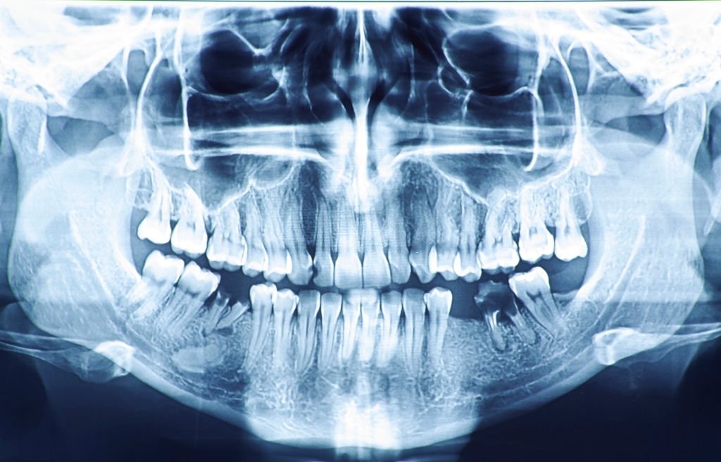
Panoramic X-rays– captures the entire mouth region on a single x-ray. Panoramic x-rays are useful in detecting fully erupted, newly emerged, impacted teeth, and helps diagnose tumors.
Dental Computed Tomography (CT scanning)- is a type of imaging that looks at interior structures in 3-D (three dimensions). This type of imaging is used to detect cysts, tumors or fractures. They also help evaluate bone strength for the placement of dental implants and difficult extractions.
Cone Beam CT– is a type of imaging that provides high quality 3D images of the soft tissue, nerves, and bone. This type of imaging acts as a guide for dental implant placement, as well as can detect any complications in the gums, tooth root, and jaws.
What are the benefits of low radiation digital radiography?
Aside from the physical and environmental impacts of traditional x-rays, digital x-rays reduces radiation exposure by up to 90% making it a safe, precise and effective imaging methodology. At Complete Dental Care of Avon we use a digital x-ray technology that leverages ALARA(As Low As Reasonably Achievable) principle to obtain digital x-rays. This ensures that you receive the minimum dose of radiation which is within the annual mean dose range. When performed with the proper precautionary techniques, there is very little cause for concern.
Furthermore it reduces the wait-time of our patients drastically.
10740 E US Hwy 36, Avon, IN 46123
317-271-3079
Office Hours
Monday 7:00 AM to 4:00 PM
Tuesday 7:00 AM to 4:00 PM
Wednesday 7:00 AM to 4:00 PM
Thursday 7:00 AM to 4:00 PM
Friday By Appointment